
Dermatological Ultrasound
- Patrick Catricala

- Mar 19
- 3 min read
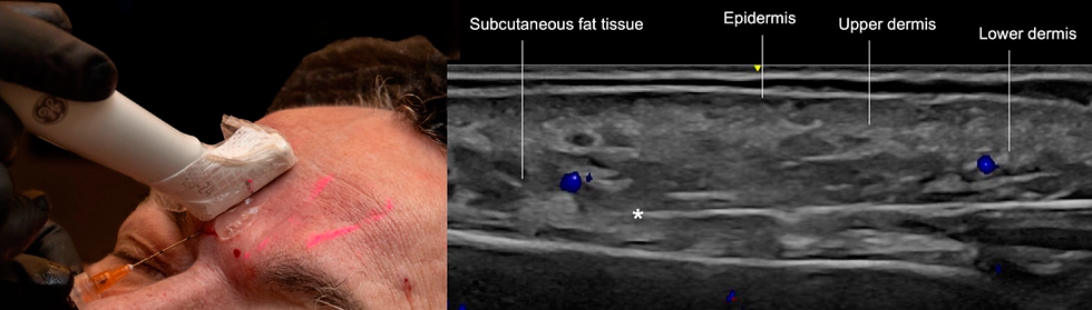
Dermatological ultrasound is a non-invasive, high-resolution imaging tool that has become indispensable in the evaluation and monitoring of injectable products used in cosmetic medicine.
It allows for detailed visualization of the skin layers and subcutaneous tissues, enabling precise identification of the location, quantity, and even the composition (in some cases, differentiating fillers from edema or granulomas) of the injected materials.
This is crucial not only for planning new procedures, ensuring safe and effective application, but also for the early detection of potential complications, such as product migration, inflammatory reactions, nodule formation, or infections.
With ultrasound, healthcare professionals can monitor the integration of the product with the patient's tissues over time, optimizing results and offering greater safety in aesthetic treatments.
Dr. Patrick Felipe Catricala
Examination performed by Dr. Patrick Felipe Catricala , specialist in diagnostic imaging with subspecialization in musculoskeletal imaging.
Dr. Patrick Catricala is a radiologist with extensive dermatological knowledge. He teaches in the Radiology and Diagnostic Imaging residency program at the Federal University.
from São Paulo.
There was interest in learning due to questions and evaluation of results from his wife, a dermatologist ( Dr. Flávia Catricala ).
Exam cost
It varies depending on the region.
Dermatological Ultrasound
How long does a dermatological ultrasound take?
The examination is estimated to last 20 to 30 minutes.
Dermatological ultrasound can be a useful tool for evaluating the effectiveness of dermal fillers.
Ultrasound is a non-invasive examination that uses high-frequency sound waves to create images of the inside of the body.
When used on the skin, it can reveal the inner layers of the skin, including subcutaneous tissue and deeper structures.
When evaluating dermal fillers with ultrasound, the doctor can see if the product has been distributed evenly and if it has reached the appropriate layers of the skin.
It is also possible to assess whether the product has been reabsorbed over time. This can help determine if any specific treatment is needed or if a new application of the product is possible.
Dermatological Ultrasound to Identify Hyaluronic Acid
Dermatological ultrasound can be used to assess the presence of hyaluronic acid in the hypodermis, the deepest layer of the skin, where dermal fillers are usually injected.
Ultrasound can also be used to assess the distribution of hyaluronic acid in the hypodermis and to check if the product has been injected uniformly. In addition, ultrasound can show if there has been any reabsorption of hyaluronic acid over time, which can be important in determining if touch-up sessions are needed.
Furthermore, it is possible to assess complications, the main one being reactive granulomas forming nodules in the hypodermis, many of which are palpable and tend not to reabsorb spontaneously.
Hyaluronidase is an enzyme that can be used to dissolve hyaluronic acid. When a dermal filler is applied excessively, unevenly, or with unsatisfactory results, hyaluronidase can be used to correct these problems.
Dermatological ultrasound can be used to help guide the injection of hyaluronidase into specific areas of the dermal filler, thus improving the effectiveness and precision of the treatment.


Dermatological Ultrasound to identify - Lifting Threads
Dermatological ultrasound can be used to evaluate the effectiveness of thread lifts, a cosmetic procedure that uses absorbable or non-absorbable threads to lift and support the skin. Ultrasound can show the position of the threads in the skin and assess whether they are properly positioned.
Ultrasound can also be useful for assessing thread reabsorption and determining if adjustments or additional sessions are needed.
Furthermore, it is possible to assess potential complications inherent to the procedure, such as infections, vascular injuries, or even malpositioning of the threads.
Bibliographic reference:

Comments